 Invoice
Knee pain – causes, muscles involved and treatment options
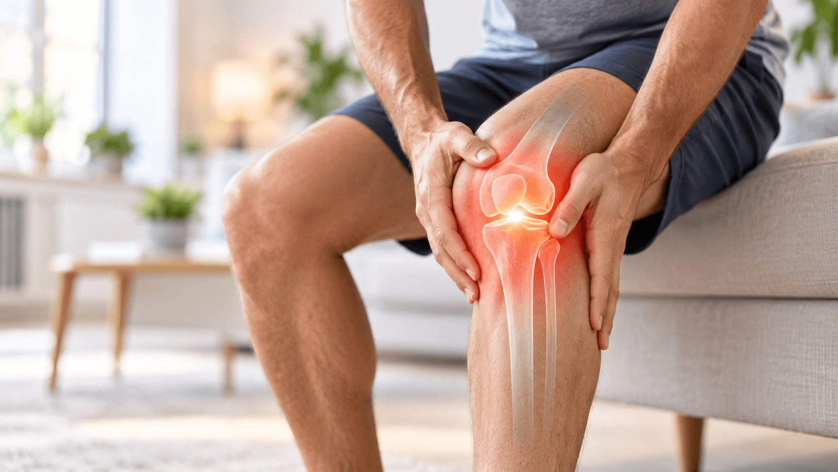
Knee pain does not always mean an injury. It is often the result of overload: pain around the kneecap, an irritated tendon or tight muscles in the thigh and calf. On this page we explain the most common non-traumatic causes, the muscles involved and what we can help with at MV Therapy – and when you should see a doctor or orthopaedist.
When to see a doctor, orthopaedist or call emergency services (112)
- An acute injury (fall, blow, twist) followed by swelling of the knee within hours
- You cannot bear weight on the leg or the knee is visibly deformed – call 112 or go to the emergency department
- The knee locks (cannot fully straighten or bend) or gives way when walking
- Suspected ligament or meniscus tear: a pop at the time of injury, instability, recurring swelling
- A hot, red and swollen joint (especially with a joint replacement) – go to the emergency department the same day or call 112.
- Swelling, pain and warmth in the calf (possible thrombosis) – see a doctor immediately; if you have shortness of breath or chest pain, call 112 immediately.
- Night pain or rest pain that does not ease, unexplained weight loss, or knee pain in a child/adolescent without a clear cause
Types of knee pain and common causes
Acute and chronic pain
Acute pain starts suddenly, usually after an injury, and first needs a doctor's assessment. Chronic or overuse pain develops gradually over weeks or months and is related to load, movement patterns and tissue capacity.
Patellofemoral pain syndrome
Pain around or behind the kneecap that worsens with squatting, stairs, running and prolonged sitting with bent knees. One of the most common causes of knee pain in active people.
Jumper's knee (patellar tendinopathy)
Pain just below the lower edge of the kneecap, especially during jumping, squatting and going down stairs. Quadriceps tendinopathy gives similar pain above the upper edge of the kneecap.
Runner's knee (iliotibial band syndrome)
Burning or sharp pain on the outside of the knee that typically appears after a certain running distance and worsens when running downhill.
Pes anserine pain
Pain and tenderness on the inner side of the knee just below the joint line, where the tendons of three thigh muscles attach. Seen both in runners and in people with knee osteoarthritis.
Early knee osteoarthritis
Joint stiffness in the morning or after sitting, pain with load and sometimes crepitus. The diagnosis is made by a doctor; manual therapy helps ease pain and stiffness and supports mobility.
Muscles and structures involved in knee pain
The knee sits between two long levers, so the condition of the hip and calf muscles directly affects knee load. Referred pain patterns are based on Travell and Simons' trigger point manual and are clinical descriptions, not a diagnosis.
Quadriceps: vastus medialis
Helps stabilise kneecap movement. Trigger points may refer pain to the front and inner knee; Travell and Simons also link them to sudden buckling of the knee.
Vastus lateralis
The largest part of the quadriceps. Trigger points refer pain to the outer thigh and outer edge of the knee; tension may affect kneecap mobility.
Rectus femoris
Crosses both the hip and knee joints. Trigger points refer pain around and behind the kneecap, often at night.
Tensor fasciae latae and the iliotibial band
The muscle tensions the iliotibial band, which runs down the outer thigh to the knee. Trigger points in the muscle may refer pain down the outer thigh to the knee; the band itself is tendinous tissue, not muscle.
Hamstrings
Bend the knee and control rotation of the lower leg. Biceps femoris trigger points refer pain to the back of the knee; the semitendinosus tendon is part of the pes anserinus.
Calf muscle (gastrocnemius)
Attaches to the lower end of the thigh bone. Trigger points in the medial head may refer pain to the back of the knee and the foot.
Popliteus
A small deep muscle that "unlocks" the straight knee. Causes pain at the back of the knee, especially when crouching, walking downhill or running.
Gluteus medius
Controls the pelvis and thigh when standing on one leg. It does not refer pain to the knee, but weakness may increase knee load; reducing its tension and improving mobility helps distribute the load on the knee more evenly.
Why overuse pain develops
Load and capacity. Most often, load (running distance, jumps, squats, stairs, body weight) has increased faster than the tissues can adapt. That is why sensible load management is the basis of every treatment plan.
Hip and foot control. People with kneecap pain may show changes in how the hip and knee muscles work: some muscles are overloaded and tight, while others do not support the knee enough. Muscle work helps balance muscle tension and reduce the load on the knee.
An overloaded tendon. A tendon irritated by repeated load becomes painful and sensitive, especially with jumping, squatting and stairs. The right load together with targeted treatment helps the tendon recover – an irritated tendon needs reduced load, not complete rest.
Muscle tension and trigger points. Tight thigh and calf muscles with trigger points can refer pain around the knee and restrict movement. Treating the muscles may reduce pain and improve mobility.
How our methods may help
Medical massage
Reducing tension in the thigh, hamstring and calf muscles. Massage may ease pain, improve muscle elasticity and knee mobility, and reduce the load on the knee.
Trigger point therapy
Targeted work on trigger points in the quadriceps, tensor fasciae latae, popliteus and calf when they refer pain around the knee.
Manual therapy and joint mobilisation
Improving mobility of the knee joint and kneecap, especially when stiff. Knee joint mobilisation reduces pain and improves knee function.[1]
Shockwave therapy (ESWT)
Shockwave therapy reduces chronic patellar tendon pain (jumper’s knee) and improves knee function. According to a systematic review, it was more effective than other non-operative treatments in several studies[2]. Usually 3–6 sessions at 4–7-day intervals.
High-intensity magnetic therapy (EMTT)
We use it to ease muscle tension and pain in knee osteoarthritis and tendon problems, often together with manual therapy.
Kinesio taping
May reduce kneecap pain and make movement easier[3]. The tape usually stays on for 3–5 days.
Therapeutic sports massage
For runners and jumping athletes: recovery of the outer and back thigh and calf muscles alongside training load.
What the research says
Below is a short summary of the systematic reviews and meta-analyses that support the methods we use.
Mobilisation reduces knee pain
In a review of 12 randomised trials, knee joint mobilisation reduced pain and improved knee function[1].
Shockwave for patellar tendon pain
According to a systematic review, shockwave therapy reduced chronic patellar tendon pain and improved function, and in several studies it was more effective than other non-operative treatments[2].
Taping for patellofemoral pain
In a meta-analysis of 10 randomised trials, kinesio taping reduced kneecap pain and improved knee function[3].
Manual therapy for knee osteoarthritis
According to a meta-analysis of 14 trials, manual therapy for knee osteoarthritis reduced pain and stiffness and improved function[4].
Treatment course step by step
1. Assessment and checking for red flags
We ask how the pain started, about load and previous injuries, assess mobility and muscles and check contraindications. If we suspect an injury, joint inflammation or another condition that needs a doctor, we refer you to a doctor or orthopaedist.
2. Reducing pain and tension
During active treatment, sessions are 4–7 days apart: massage, trigger point work, and where needed mobilisation, taping or device-based therapy. At the same time we adjust your load.
3. Gradual return to load
As pain eases, we continue the muscle work and support a gradual return to your usual load – running, jumping or stairs.
4. Maintenance and prevention
After active treatment, regular maintenance sessions help keep the result and prevent flare-ups; appointments can be arranged as needed.
What you can do yourself
- Temporarily reduce the load that provokes pain (e.g. running volume, deep squats, jumps), but keep moving – complete rest is usually not needed.
- A simple rule: mild discomfort during training is acceptable, but pain should not be worse the next morning than before.
- Increase running volume gradually and note when and where the pain appears.
- When sitting for long periods, stand up from time to time and straighten your legs.
- If pain worsens, swelling appears or the knee locks, reduce the load and see a doctor.
Does your knee hurt under load?
Book an appointment at Viimsi Hospital (Ravi tee 4) or in Pärnu (Rüütli 47). We assess mobility and muscles and let you know if a doctor's assessment is also needed.
Frequently asked questions
Related services and topics
References
- Jayaseelan DJ, Scalzitti DA, Palmer G, Immerman A, Courtney CA. The effects of joint mobilization on individuals with patellofemoral pain: a systematic review. Clin Rehabil. 2018;32(6):722-733. doi.org/10.1177/0269215517753971
- Mani-Babu S, Morrissey D, Waugh C, Screen H, Barton C. The effectiveness of extracorporeal shock wave therapy in lower limb tendinopathy: a systematic review. Am J Sports Med. 2015;43(3):752-761. doi.org/10.1177/0363546514531911
- Jiao H, Tao M, Cui X. Efficacy on pain and knee function of Kinesio taping among patients with patellofemoral pain syndrome: a systematic review and meta-analysis. BMC Musculoskelet Disord. 2025;26(1):388. doi.org/10.1186/s12891-025-08627-7
- Xu Q, Chen B, Wang Y, Wang X, Han D, Ding D, Zheng Y, Cao Y, Zhan H, Zhou Y. The effectiveness of manual therapy for relieving pain, stiffness, and dysfunction in knee osteoarthritis: a systematic review and meta-analysis. Pain Physician. 2017;20(4):229-243. pubmed.ncbi.nlm.nih.gov/28535547/
This page is for information only and does not replace a doctor's diagnosis or treatment. In case of acute injury, swelling, joint locking or signs of inflammation, see a doctor.